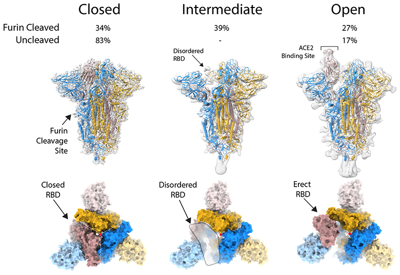
Figure 1. Structure of protease-cleaved SARS-CoV-2 spike glycoprotein.
Three structures are calculated from micrographs of furin-cleaved material: closed, intermediate and open forms of approximately equal proportions. In the uncleaved material, most of the population represents the closed form with a small proportion in the open conformation. Density maps for the three types of particles, overlaid with a ribbon representation of the built molecular models, as viewed with the long axis of the trimer vertical (middle panel); the three monomers are coloured blue, yellow and brown. An orthogonal view (lower panel) looking down the long axis (indicated by a red dot), the colouring is as in the middle panel with the NTDs in a lighter hue.

