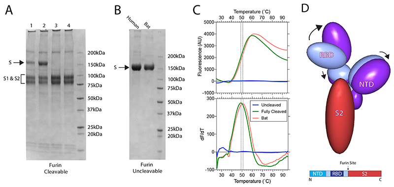
Extended Data Fig. 1. Biochemical analyses of spike proteins.
(A) SDS-PAGE of furin-cleavable SARS-CoV-2 protein. 1: 5 hr furin cleavage; 2: Expressed protein, not cleaved in vitro; 3: 12 hr furin cleavage; 4: 32 hr furin cleavage. (B) SDS-PAGE of uncleavable bat and human virus S proteins. (C) Differential Scanning Fluorimetry measurement of melting temperature for uncleavable human and bat, and fully-furin-cleaved SARS-CoV-2 proteins. (D) The changes in domain orientation, between the closed and open forms, shown schematically, for the monomer that undergoes the most substantive change in the RBD position. The image is produced by the CCP4 MG 'bloboid representation' and is calculated from the shape and centre of mass of the molecular model. Also shown is a bar representation of the domains with the furin cleavage site indicated.

