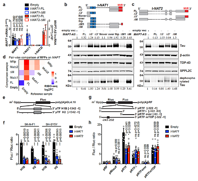
Fig. 3. MAPT-AS1 controls tau translation through embedded inverted MIR.
Stable expression in SH-SY5Y cells a, MAPT-AS1 and MAPT expression by qRT-PCR (2–ΔΔCt); Empty vector (Empty), full-length or mutant t-NAT1 (t-NAT1FL; t-NAT1ΔM), or t-NAT2l (t-NAT2FL; t-NAT2ΔM), MIR deletion (ΔM) (mean±s.e.m., n=6, 3 clones in 2 experiments, one-way ANOVA with Dunnett’s test), t-NAT1 (b) and t-NAT2 (c) with: full-length (FL), 5’-deletion (Δ5’); 3’-deletion (Δ3’); regions not-overlapping (Nover) or overlapping (over) with MAPT 5’UTR; flipped overlapping region (flip); partial (ΔM1) or full MIR deletion (ΔM). AS-region overlapping MAPT 5’UTR in blue; chevrons indicate orientation. t-NAT1-FL (b), t-NAT2-FL (c) reduce endogenous total- and dephosphorylated-tau (λ-phosphatase), suggesting regulation is independent of tau phosphorylation. Inverted MIR (red) is essential for controlling tau levels. Numbers above total-tau and below dephosphorylated-tau indicate levels normalised to β-actin, TDP-43 and SPPL2C geometric mean. d, Pairwise comparison heatmap of RIBO-seq ribosome footprints (RFPs) along MAPT from 3 independent SH-SY5Y clones expressing Empty-vector, t-NAT1 (FL), deletion of MIR motif-1 (MutΔ1) or motif-2 (MutΔ2) as in Fig.4a, MIR deletion (ΔM), MIR flipped (Mflip). FL significantly decreases MAPT RFPs compared to Empty (log2FC=-1.45, p=0.036, Wald test with Bonferroni correction). e, pTF reporters: a 1,342 nt genomic fragment spanning MAPT promoter, 5’UTR (grey box) and intron segment, upstream to firefly luciferase (Fluc) ORF. Haplotypes H1B and H2, (7 SNPs), were tested. f, FL t-NAT1 and t-NAT2 transient expression significantly repress Fluc translation normalised to Renilla luciferase (mean±s.e.m., one-way ANOVA with Dunnett’s test, n=3 SK-N-F1, n=6 SH-SY5Y independent experiments). g, Bicistronic reporters: MAPT 5’ UTR inserted between Renilla (Rluc) and Fluc ORFs in pRF vector62, resulted in pRTF. Truncations (pRTFΔ and pRTFover) or 5’TOP motif mutation (pRTFmTOP) reduced tau-IRES activity. Hepatitis C virus IRES (pRhcvF), positive control. h, SH-SY5Y cells stably expressing empty vector (Empty), t-NAT1 or t-NAT2, were transfected with constructs in (g) and capindependent translation (Fluc/Rluc ratio) measured. Control cells (Empty) transfected with pRTF showed a ~15- fold increase in Fluc/Rluc ratio over negative control pRF vector, and a ~3.7-fold increase over pRhcvF; FL t-NAT1 or t-NAT2 expression significantly reduced tau-IRES activity. (n=3 SH-SY5Y clones in 2 independent experiments, mean±s.e.m., two-sided Kruskal-Wallis with Dunn’s test).

