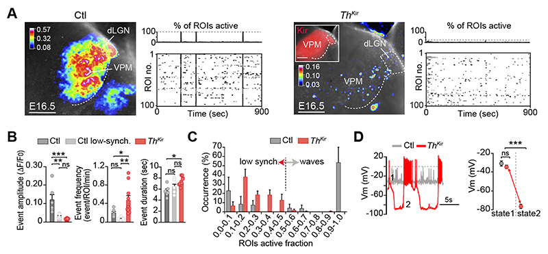
Fig.2. Desynchronizing the embryonic thalamic pattern of activity.
(A) Maximal projection of ex vivo spontaneous calcium activity in the ventral postero-medial nucleus (VPM) and accompanying raster plots in control and ThKir slices at E16.5. (B) Properties of the VPM calcium events (n = 6 control, n = 10 ThKir; *P < 0.05, **P < 0.01, ***P < 0.001). (C) Percentage distribution of active ROIs. (D) Representative traces and quantification of membrane potential (Vm) in control and ThKir neurons recorded at E16.5-E18.5 (control n = 7; ThKir n = 7). ***P < 0.001. dLGN, dorso-lateral geniculate nucleus. Scale bars, 200 μm. Data are means ± SEM.

