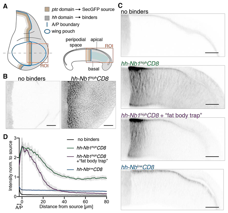
Fig. 1. Establishment of a GFP gradient in a developing epithelium.
(A) Schematic representation of a wing imaginal disc of Drosophila. SecGFP is expressed under the control of the ptc promoter (brown), while a membrane-tethered anti-GFP nanobody is expressed under the control of the hh promoter (grey). ROI indicates the areas depicted in panels B and C. Blue shading indicates the region used to generate the profiles shown in D. (B) In the absence of binders, SecGFP can be seen at the source but is not detectable in basolateral focal planes. Upon expression of high-affinity binders in the posterior compartment (hh-Nb1highCD8), a gradient is readily seen. (C) Cross-sections of imaginal discs expressing SecGFP in the ptc domain, showing that, in the absence of binders, GFP is detectable in the peripodial space but not in the basolateral space. In the presence of binders (hh-Nb1highCD8), a gradient can be seen in the basolateral space but with a non-zero tail, which is largely abrogated by concomitant activation of UAS-Nb1highCD8 in the fat body (+ “fat body trap”). Only a shallow basolateral gradient is detected when a low-affinity binder is expressed (hh-NblowCD8). (D) Fluorescence intensity profiles derived from preparations like those shown in panel C. The vertical dotted line marks the estimated posterior edge of the source. The numbers of discs analyzed are as follows: No binders, n=10; hh-Nb1highCD8, n=11; hh-Nb1highCD8 + fat body trap, n=7; hh-NblowCD8, n=10. Scale bars: 20 μm

