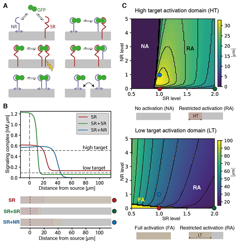
Fig. 5. Modeling the effect of GPI-anchored non-signaling receptors on gradient length scale.
(A) Schematic representation of the molecular interactions considered by our model, including GFP dimer handover and NR hopping. Signal transduction (yellow lightning bolt) is activated by GFP-bound signaling receptor dimers (SR). See Supplementary Theory for details. (B) Predicted profiles of signaling complexes in three conditions: a reference case with signaling receptors only (SR; red), doubling SR levels (SR + SR; green), and adding non-signaling receptors (SR + NR; blue). As observed experimentally, doubling SR leads to a steeper gradient, while adding NR reduces GFPhemo signaling and extends the gradient, due to non-signaling receptor effective diffusion. For illustration, arbitrary thresholds are chosen to indicate the position where high- and low-level target genes would be activated (see Supplementary Tables 2 and 3 for parameter values). (C) Width of the high (top) and low (bottom) target activation domain (arbitrary threshold shown in panel B), as a function of normalized levels of SR and NR. Warmer colors indicate a wider target activation domain. Colored dots show parameter combinations used in panel B. Top: For the normalized SR value of 1, increasing NR initially lengthens the high target domain, while further increase shortens it by preventing access of GFP to SR (as observed experimentally, Fig. S11). Bottom: For the normalized SR value of 1 and in the absence of NR, GFPhemo signaling dominates and low target gene is activated throughout (bright yellow region). Increasing SR or NR production both lead to a reduction in the low target domain size.

