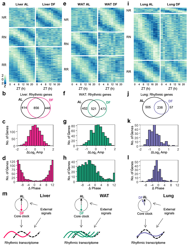
Figure 2. Daytime-restricted feeding affects the rhythmic transcriptome in a tissue-specific manner.
Rhythmic transcriptome analyses of (a-d) Liver, (e-h) White Adipose Tissue (WAT), and (i-l) Lung from mice fed either ad libitum (AL) or exclusively during the light-phase (DF) for 30 days. (a, e, and i) Heatmap of expression profiles of genes that were non-rhythmic in AL and rhythmic in DF (NR, Qmin <0.05, PHR,AL>0.05, PHR,DF≤0.05, JTK_CYCLE and Harmonic Regression, see also Methods section); genes that were rhythmic in AL and non-rhythmic in DF (RN, Qmin <0.05, PHR,AL≤0.05, PHR,DF>0.05); and genes that were rhythmic in both (RR, Qmin <0.05, PHR,AL≤0.05, PHR,DF≤0.05). Data is presented as z-scores of the average expression in each ZT. (b, f, and j) Venn diagrams representing the overlap between rhythmic genes in AL or in DF. (c, g, and k) Histogram representing the distribution of amplitude-differences between DF and AL, of the common rhythmic genes in both conditions. (d, h, and l) Histogram representing the distribution of phase differences between DF and AL for the common rhythmic genes in both conditions. (m) Graphical depiction of the main findings. The transcript rhythmicity is driven both by the tissue’s internal core clock and by various external signals. Upon DF, which affects external rhythmicity, the core clock in the different tissues is differentially phase-shifted. The effect on the rhythmic transcriptome is tissue-specific as well: the liver transcriptome is phase-inverted, the lung transcriptome is mildly affected, while the WAT transcriptome exhibits wide phase-shift distribution.

