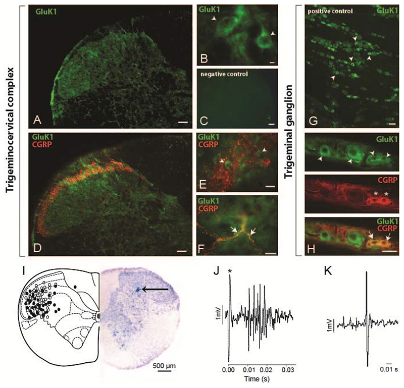
Figure 1. GluK1-like immunofluorescent staining within the trigeminocervical complex and trigeminal ganglia, and reconstruction of recording sites.
A-B. Photomicrographs of the trigeminocervical complex taken from the C1level, showing GluK1-like staining (green). Cell bodies and punctate staining was evident in laminae I-III and mainly cell bodies were seen in deeper laminae. Stained cells were round or pear-shaped. D-F. Within lamina I and outer lamina II, where calcitonin gene-related peptide (CGRP)-like fibers project (red), GluK1-like staining (green) was seen in cell bodies as well as in punctate staining, which might represent proximal processes or GluK1 primary fibers as some co-localization (yellow) with CGRP was seen (Figure 1G). C. A photomicrograph of a negative control section of the TCC. G. A photomicrograph of GluK1-like cells (green) within the trigeminal ganglion. H. GluK1-like cells (green) mainly co-localized with small size CGRP-positive cells (red) within the trigeminal ganglion. Arrowheads present examples of GluK1-like positive cells, stars indicate CGRP-like positive cells and arrows show co-localization of CGRP- and GluK1-like fibers (F) or cells (H). Scale bars = 100 μm (A, C, D), 50 μm (E, G, H), 10 μm (B, F). I. Reconstruction of recording sites within the C1 spinal cord level, plotted after Paxinos and Watson [43], identified histologically (solid circles represent pontamine sky blue spots) and by microdrive readings (open circles). A photomicrograph demonstrating an original recording site marked by ejection of pontamine sky blue (arrow) is shown. J. An original trace showing a cluster of cells in the trigeminocervical complex, firing in response to stimulation of the middle meningeal artery (100 μs, 0.5 Hz, 12 volts; * stimulus artefact). K. Original tracing from a neuron in the trigeminocervical complex responding to microiontophoresis of L-glutamate.

