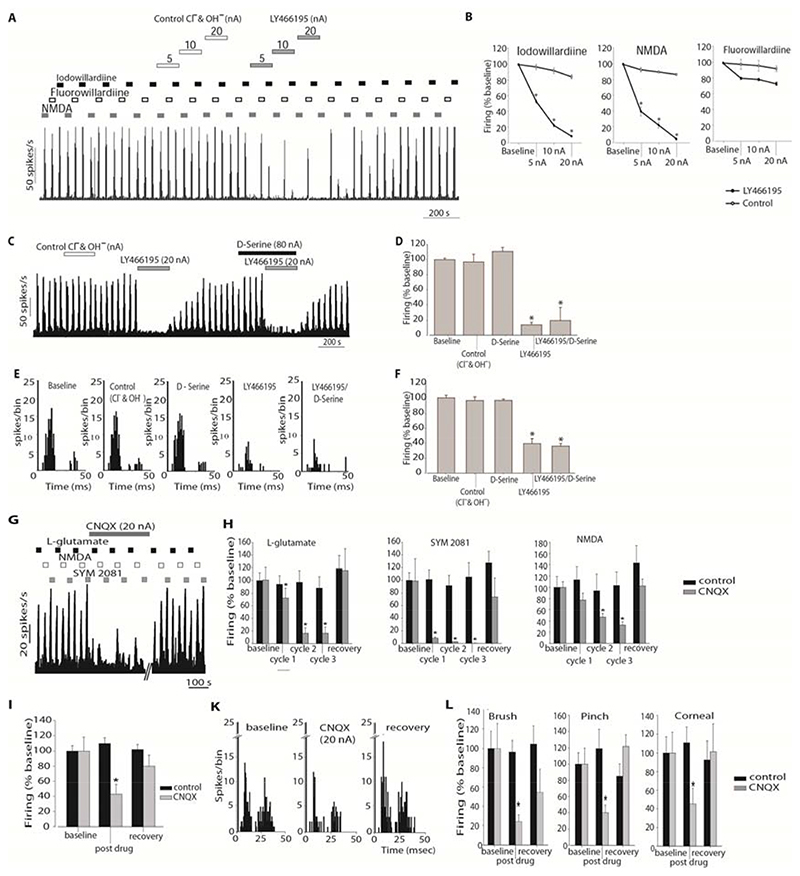
Figure 3.
A. Example of the effects of LY466195 on the firing rates to pulsed ejections of Fluorowillardiine, Iodowillardiine and NMDA. Cell firing in response to the ejected agonists returned to baseline levels within 2-5 minutes after LY466195 microiontophoresis ceased. B. Comparison of the effect of LY466195 and control ions (Cl-, OH-) on iodowillardiine, NMDA and fluorowillardiine evoked responses. Cell firing in response to iodowillardiine and NMDA was significantly inhibited by microiontophoretically administered LY466195. C. Example of the response of a trigeminocervical neuron to pulsed NMDA. Ejection of serine, LY466195 and control ions are shown with the solid bars. Ejection of LY466195 demonstrated a potent inhibitory effect on NMDA-evoked firing that was not reversed by pre-treatment with serine. D. Comparison of the NMDA evoked firing during ejection of control ions and LY466195. E. Post-stimulus histograms generated from a representative trigeminocervical neuron following electrical stimulation of the middle meningeal artery. Microiontophoresis of serine or control ions at the same current had no effect on neuronal firing. Co-ejection of serine with LY466195 failed to block the inhibitory actions of LY466195. F. Comparison of the middle meningeal artery stimulation-evoked firing under the influence of LY466195, serine and control ions. G. Example of the effects of CNQX on the firing rates to pulsed ejections of the L-glutamate, Iodowillardiine and SYM 2081. Cell firing in response to the ejected agonists returned to baseline levels within 30 min after CNQX microiontophoresis ceased. B-H. Effects of CNQX on cell firing in response to L-glutamate, SYM 2081 and NMDA, separately for each ejection cycle. CNQX strongly inhibited L-glutamate, SYM 2081 and NMDA-evoked firing. Ejection of control ions had no significant effects on glutamate agonists-evoked firing. I. Comparison of the middle meningeal artery stimulation-evoked firing under the influence of CNQX and control ions. CNQX significantly inhibited neuronal firing in response to trigeminovascular stimulation. K. Post-stimulus histograms generated from a representative trigeminocervical neuron following electrical stimulation of the middle meningeal artery during baseline conditions, microiontophoresis of CNQX and recovery. neuronal firing. L. Effects of microiontophoretically delivered CNQX on the firing rates of second order neurons in response to receptive field characterisation. Microiontophoretic application of CNQX at 20 nA significantly inhibited firing to both innocuous (brush) and nocuous (corneal brush; pinch) stimulation of the ophthalmic dermatome, while a lower ejection current (10 nA) significantly inhibited noxious stimulation of the receptive field. * P < 0.05 compared to control

