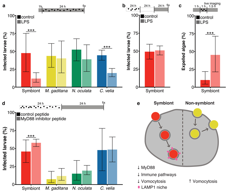
Fig. 5. Immune stimulation enhances expulsion of symbionts during initial interaction.
a Percentage of symbiont-infected Aiptasia larvae was significantly (p = 0.0000000024) reduced when larvae were pre-treated for 1 hour with LPS, followed by a 24-hour exposure to microalgae and LPS. Percentage of C. velia-infected larvae was also reduced (p = 0.00000000024), but to a lesser degree; there was no effect observed for M. gaditana- or N. oculata-infected larvae. (n=5) b LPS exposure for 24 hours after established symbiosis (24 hours infection + 24 hours incubation without symbionts) did not influence symbiont maintenance. (n=5) c LPS treatment induced expulsion. LPS exposure for 1 hour before and during 1 hour of infection, significantly (p = 0.00038) enhanced the number of larvae with expulsion events as observed during 1.5 hours of live imaging. For the LPS treatment n=3 with total 42 microalgae; for the control n=4 with total of 50 microalgae. d MyD88 inhibition enhanced maintenance of symbionts in larvae (p = 0.0000053), while maintenance of other microalgae was unaffected. For the symbionts n=10; for M. gaditana n=3; for N. oculata n=13; and for C. velia n=3. e Model of microalgae uptake in Aiptasia endodermal cells. While uptake of symbionts (red) leads to downregulation of immunity genes until a functional LAMP1-positive symbiosome is formed, non-symbiotic microalgae (yellow) do not elicit a strong transcriptomic response in the host cells and are subsequently expelled by vomocytosis. All graphs show mean ± 95 % CI with statistics based on a two-sided generalized linear mixed model accounting for repeated measurements. Bars above graphs represent duration of infection (black dots), treatment (grey fill) and live imaging (where applicable). *P < 0.05, **P < 0.01, ***P < 0.001.

