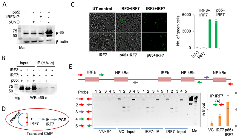
Fig. 3. NF-κB p65 interacts with IRF7 and increases its promoter occupancy.
A) Western blot (WB) showing endogenous expression of p65 in unstimulated HEK293T cells. The overexpressed TFs in different lanes are shown on the top of the blot. B) Co-IP experiment showing the physical interaction between IRF7 and NF-κB p65. The HA tags present in the IRF3 and IRF7 constructs were used to pull down the complexes before probing for p65 using a specific antibody. The blot shown is a representative of two independent Co-IP experiments. C) Bimolecular Fluorescence Complementation Assay (BiFC) shows that IRF7 interacts with p65. UT-untransfected control. The left and middle images show merged brightfield and fluorescence views; the right image is only from the fluorescence view. The quantification shown on the right graph was carried out with Kaleido (PerkinElmer). D) Schematic showing the flow of transient ChIP experiment. The p0.5kbIFNL3 construct was co-transfected along with IRF7 and ChIP was performed with anti-IRF7 antibody. E) (Top and bottom left) Standardization of primer sets for transient ChIP. After pulling down the complexes and reversing the cross-links PCR was performed with the DNA obtained using 4 different primer set combinations. The top shows the schematic of the five different IRF or NF-κB binding sites on the p0.5kbIFNL3 promoter construct. Each arrow depicts a primer. The red arrows are specific for primers entirely binding in the pGL3basic plasmid DNA sequence; the remaining arrows show primers that bind within the 0.5 kb promoter encompassing different TF binding regions. The gel picture shows the PCR products run on a 2.5% EtBr-stained agarose gel. First marker (Ma) is 1 kb ladder (GeneRuler 1 kb DNA Ladder, ready to use, Cat#SM0313,ThermoFisher Scientific, USA); second marker is 50 bp ladder (TrackIt™ 50 bp DNA Ladder, Cat#10488-043, ThermoFisher Scientific, USA). (bottom right) qPCR showing the enrichment of IRF7-bound DNA which is increased greater than 2-fold when p65 is co-transfected with IRF7. The primer set 4 was used for the qPCR (the forward primer binds downstream of NF-xBa site and the reverse primer binds within the plasmid DNA sequence). The mean values from triplicate experiments are shown, error bars depict SD. VC-vector control.

