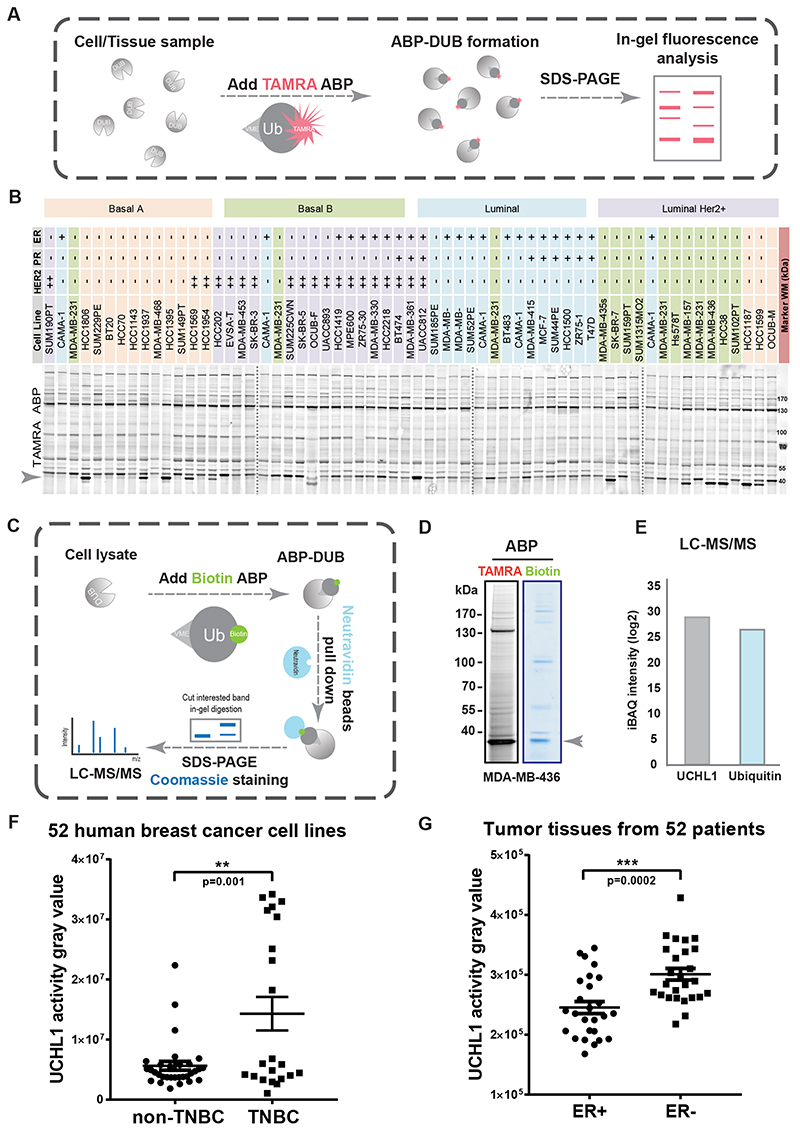
Figure 1. DUB activity profiling identified UCHL1 as being selectively highly activated in aggressive breast cancer tumor tissues and cell lines.
A, Schematic overview of DUB activity profiling with TAMRA activity based probe (ABP). B, Atlas of DUB activity in 52 breast cancer cell lines. Four gels were merged together with dashed line in between two gels. C, DUB identification workflow with Biotin ABP. D, TAMRA ABP and Biotin ABP assay in MDA-MB-436 cells. E, LC-MS/MS analysis of in-gel tryptic digestion of excised gel slice indicated in figure 1D. F, UCHL1 activity analysis of 52 breast cancer cell lines. **, P < 0.01, unpaired Student t test. G, UCHL1 activity gravy value analysis of 52 tissues from breast cancer patients. ***, P < 0.001, unpaired Student t test.

