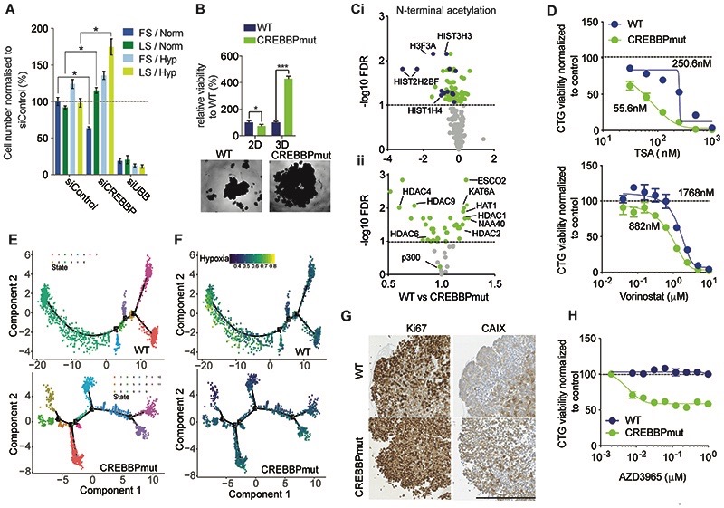
Figure 2. CREBBP loss promotes growth under nutrient stress and hypoxia and confers sensitivity to HDAC and MCT-1 inhibition.
(A) Barchart depicting normalised cell viability assessed with Cell Titre Glo of MCF10AT1 cells that were grown in 2D under differing serum (Full/10% and Low/1% FBS) and oxygen (Normoxia/20% and Hypoxia/1% O2) and in combination for four days. siUBB was used as a positive cell killing control. (B) Bar chart representing isogenic HAP1 cell growth in 2D and 3D for seven days. Representative bright-field images of WT and CREBBPmut spheroids are also shown. (C) i) Volcano plot of the significantly altered N-terminal acetylated peptides between CREBBPmut and WT cells. HAP1 WT and CREBBPmut spheroids were grown for five days. Log2 fold change is plotted against -log10 of FDR corrected p value. Histone proteins are highlighted in blue. ii) Volcano plot of fold change of total protein expression of histone acetylase and deacetylases plotted against FDR corrected p-value. (D) Dose response curves of HAP1 WT and CREBBPmut spheroids were treated with increasing concentrations of HDAC inhibitors, Tricostatin A (TSA) and Vorinostat for five days. (E) Single cell RNAseq analysis after seven days of growth. DDRTree visualization and 2-D embedding showing constructed pseudo-time transcriptional states for WT and CREBBPmut spheroid cells, depicting an increased number of branches in the CREBBPmut cells indicative of differential transcriptional programmes over time. (F) Single cell RNAseq from (E) was also analysed for the presence of a hypoxia gene signature. (G) Representative micrographs of CREBBP, Ki67, carbonic anhydrase CAIX expression in HAP1 WT and CREBBPmut spheroids. Scale bar represents 100μm, indicating increased proliferation in CREBBPmut cells and increased levels of the hypoxia marker CAIX. (H) Dose response curves of HAP1 WT and CREBBPmut spheroids treated with increasing concentrations of the selective Monocoarboxylase transporter 1 inhibitor (MCT-1) AZD3965 for 5 days, showing that CREBBPmut cells are selectively sensitive to MCT-1 inhibition. Spheroid viability was assessed using CellTiter-Glo and normalised to DMSO treated cells.

