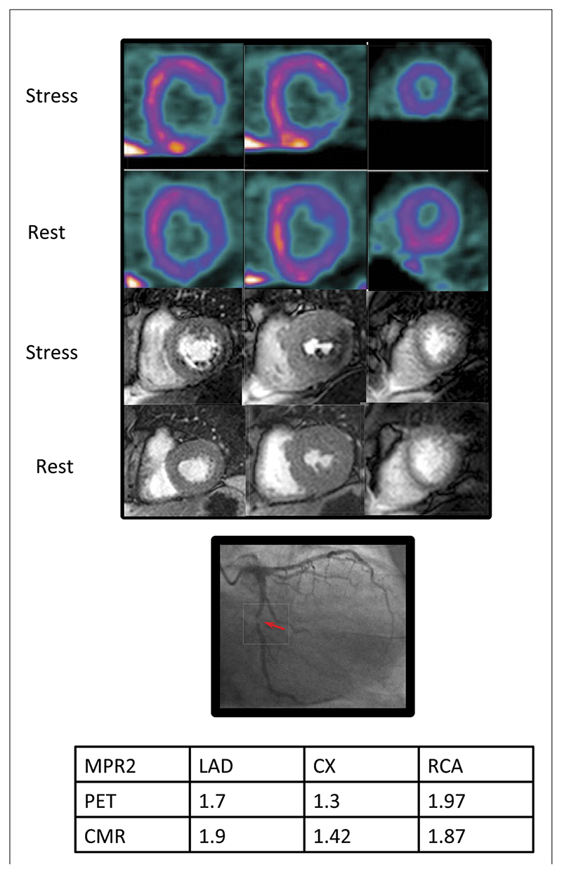
Figure 5. Case Example.
Positron emission tomography (PET) (top), cardiac magnetic resonance (CMR) (middle), and the x-ray angiogram of the left coronary artery of a 54-year-old patient with diabetes and exertional angina. Basal, mid, and apical slices have been taken from the PET study, which approximately correspond to the CMR slices. There is a stress-induced perfusion defect in the infero-lateral region from base to apex visible on both PET and CMR images. There is a corresponding severe (>95%) stenosis of the proximal circumflex artery. There was no other significant angiographic disease. Myocardial perfusion reserve of the lowest 2 segments (MPR2) for each territory are shown in the table. The MPR2 for the circumflex artery (CX) is below the cutoff of 1.44 and 1.45 for both PET and CMR, respectively. LAD = left anterior descending artery; RCA = right coronary artery.

