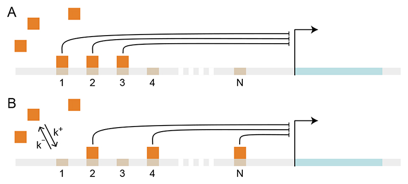
Figure 2. Schematic representation of a promoter with multiple binding sites, numbered from 1 to N.
Transcriptional repressors (orange squares) bind and unbind from binding sites (numbered platforms) at the promoter of a gene (light blue stretch). (A) and (B) illustrate two equivalent configurations of the promoter, with identical number of bound transcriptional repressors having the same inhibiting strength.

