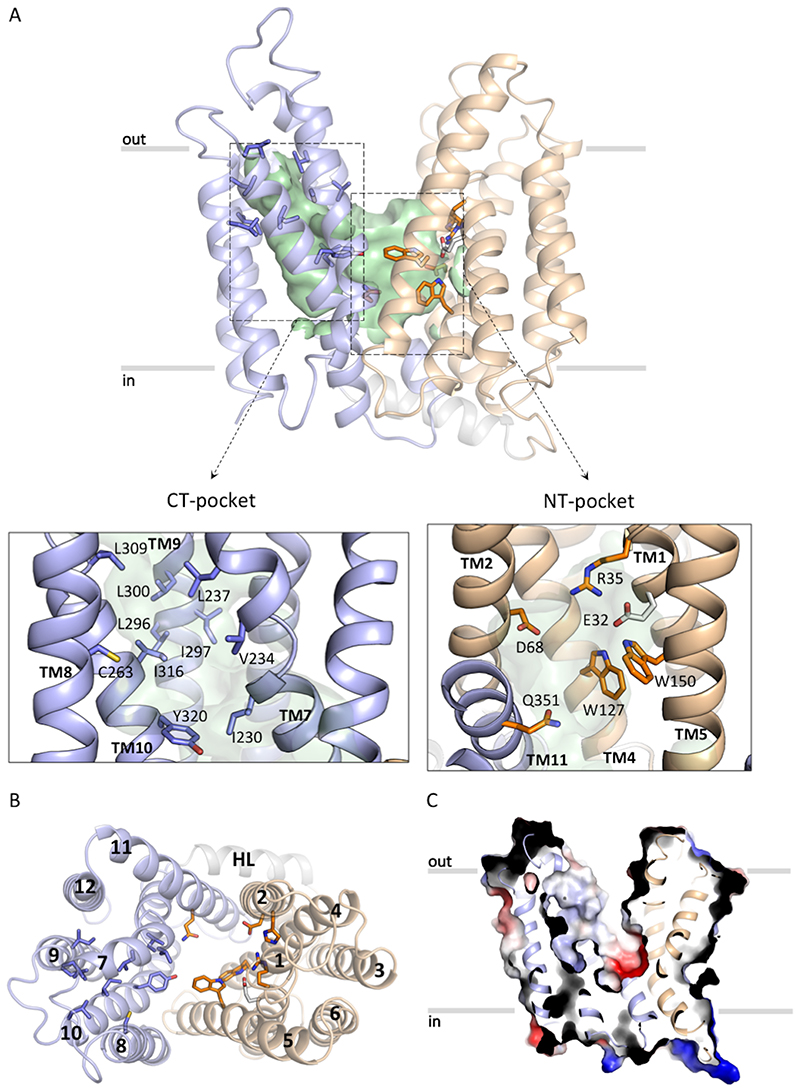
Figure 2. S. aureus LtaA structure.
A. Structure of LtaA showing its central cavity (green surface). CT: C-terminal, NT: N-terminal. The N-terminal domain is shown in light-orange, C-terminal domain is shown in light-blue. Residues forming the hydrophobic C-terminal pocket and the hydrophilic N-terminal pocket are shown. B. Top view of LtaA. Residues in sticks participate in formation of the amphiphilic cavity. C. Vacuum electrostatic surface representation of LtaA showing the internal cavity.

