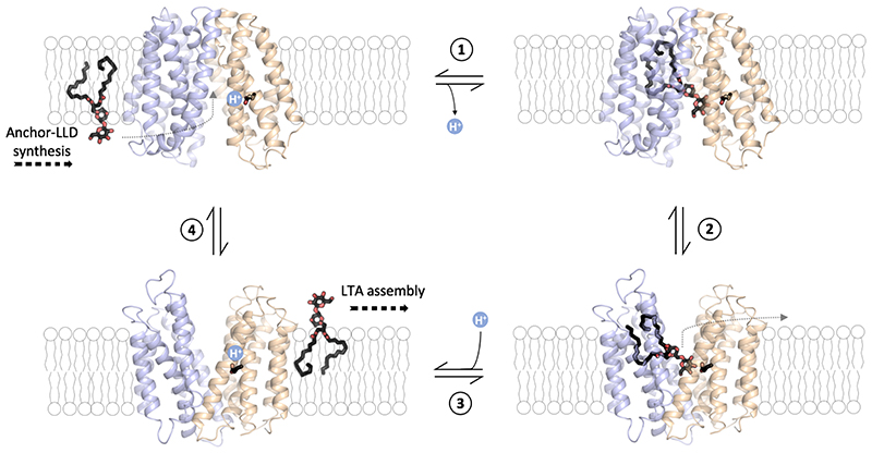
Figure 6. LtaA anchor-LLD flipping mechanism.
1 Binding of lipid-linked-disaccharide (black and red sticks) in the central cavity of LtaA in inward-facing conformation (modelled conformation) and deprotonation of E32. 2 Transition to outward-facing state (structure determined in this study). 3 Substrate release into the membrane and protonation of E32. 4 Transition to inward-facing state (modeled conformation). The N-terminal domain is shown in light-orange, C-terminal domain is shown in light-blue. The inward-facing model of LtaA was constructed by rigid body alignment of the N-terminal domain (TM1-6) and the C- terminal domain (TM7-12) to those of inward-facing LacY (PDB: 2CFQ)60.

