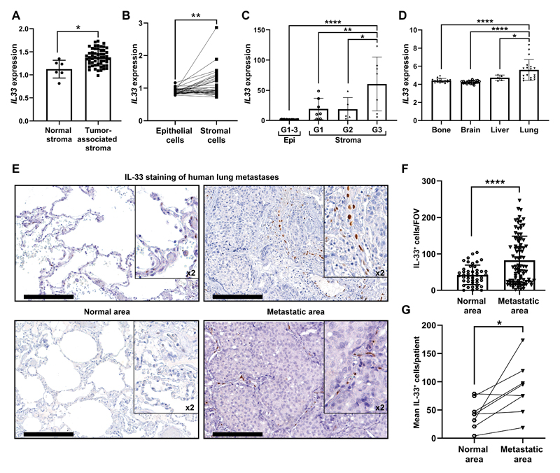
Figure 6. IL-33 is upregulated in the stroma of breast cancer primary tumors and lung metastases in human patients.
(A) IL33 expression in tumor-associated stroma as compared with normal stroma from GSE9014. Welch’s t-test, *P<0.05. (B) IL33 expression in paired patient samples of epithelial cells and tumor-associated stroma in primary breast tumors from GSE88715. Paired t-test. **P<0.01. (C) IL33 expression in primary breast-tumor tissue (Epi) and tumor associated-stroma (Stroma) from GSE14548. Stromal expression is divided into tumor grade (Stroma-grade 1: G1; Stroma-grade 2: G2; Stroma-grade 3: G3). For the epithelial expression of IL33 all grades were combined (G1-3). One-way ANOVA with Tukey’s correction for multiple comparisons, *P<0.05, **P<0.01, ****P<0.0001. (D) IL33 mRNA expression in human breast cancer metastasis from different metastatic sites. Data was derived from GSE14020. One-way ANOVA with Tukey’s correction for multiple comparisons tests. *P<0.05, **** P< 0.0001. (E) Representative IHC staining of IL-33 in lung metastases of breast cancer patients (n=8). Normal areas (remote from metastatic foci) were quantified as controls. Scale bars: 200μm. (F) Quantification of the number of IL-33+ cells per FOV in IHC performed in (c). 10-15 FOV/section. Data presented as mean ±SD; Welch’s t-test. ****P<0.0001. (G) Analysis of the mean IL-33+ cells/patient, compared with paired adjacent normal lung tissue. n=8; Paired t-test. *P<0.05.

