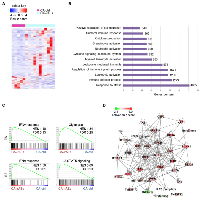
Figure 3. Shared pro-inflammatory transcriptomic profile in Tregs across different types of cancer.
Transcriptomic analysis of CD4+CD25+CD127– Tregs isolated from peripheral blood of CA-ctrl (n=5) and CA-irAEs (n=11) treated with anti-PD1. (A) Heatmap of DEGs with scaled cpm expression values (row z-score). (B) Bar plot representing pathway analysis of DEGs with adjusted p value ≤ 0.05. Each term size is listed on the end of each bar. (C) GSEA plot showing the enrichment of “IFNγ response” (NES 1.40, FDR 0.13), “Glycolysis” (NES 1.34, FDR 0.25), “IFNα response” (NES 1.59, FDR 0.01) and “IL2-STAT5 signaling” (NES 0.69, FDR 0.23) gene set. (D) Upstream regulator network from the DEGs of ‘CA-ctrl vs CA-irAEs’ using IPA analysis; red: represents activation in CA-irAEs; green: represents inactivation in CA-irAEs. For DEG analysis, the GLM statistical approach and the thresholds: |FC|≥1.5 and p<0.05 were used.

