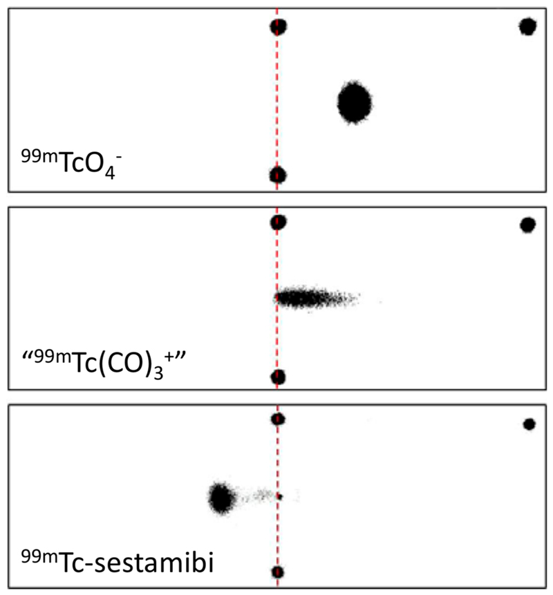
Figure 8. Phosphor images obtained from electrophoresis of [99mTc][TcO4]− (top), [99mTc]-sestamibi (middle), and “[99mTc]-[Tc(CO)3]+” in PBS (pH 7.4).
The two black spots joined by a red dashed line represent the starting point of the radioactive complexes, and the black spot at the top right indicates the position of the anode. Electrophoresis results in citrate buffer at pH 5.1, tris-HCl buffer at pH 7.4, and tris-HCl buffer at pH 8.8 are shown in SI Figure S26).

