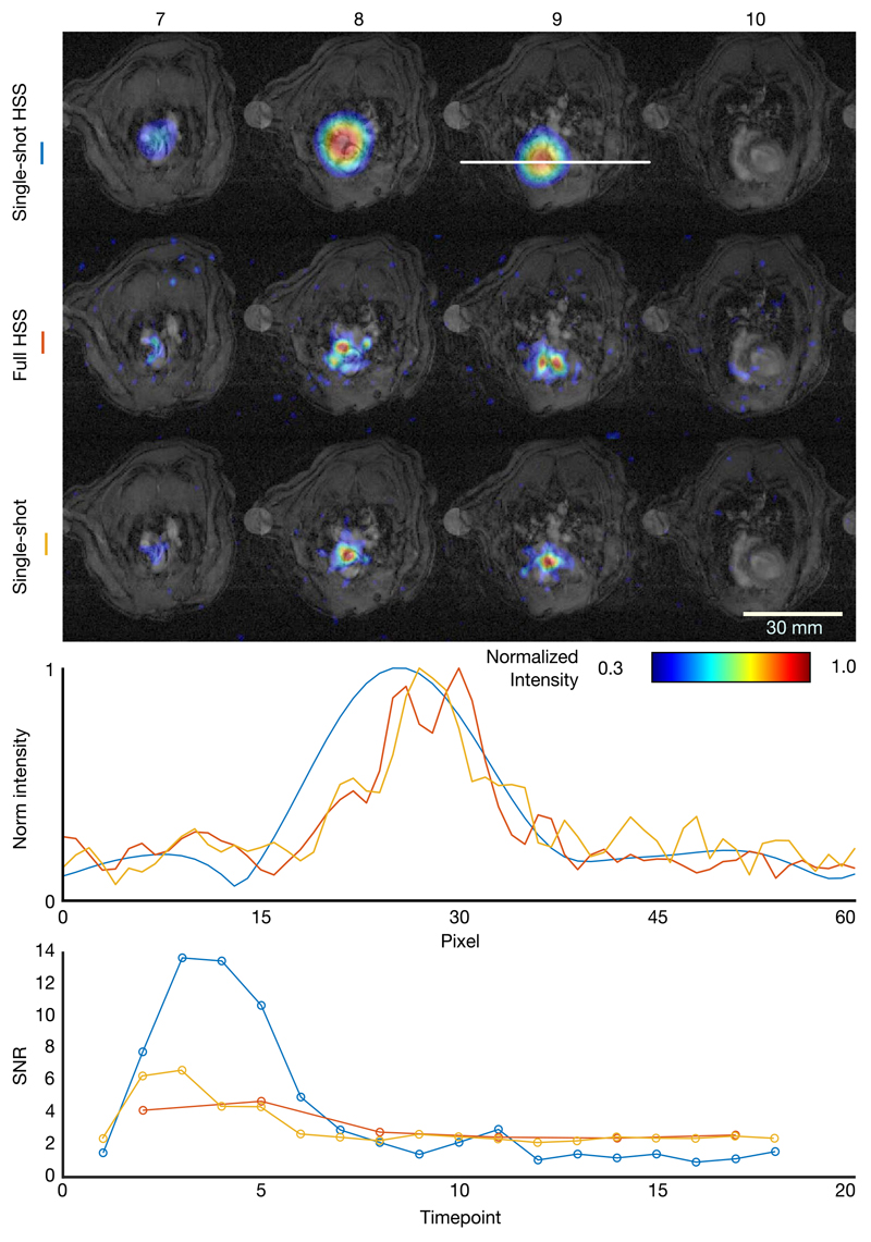
Figure 5.
Top: Axial slices 7-10 of HSS and single-shot 13C acquisitions of a rat injected with hyperpolarized [1-13C] overlaid on to matched 1H anatomical images, excited at the pyruvate resonant frequency. Middle: Line plots through slice 9 of each reconstruction in the position indicated by the white line in the single-shot HSS image. Bottom signal-to-noise ratio of pyruvate in the rat heart (Figure 5, slice 9) as a function of acquisition timepoint for single-shot and full reconstructions of a HSS acquisition in addition to a single-shot acquisition

