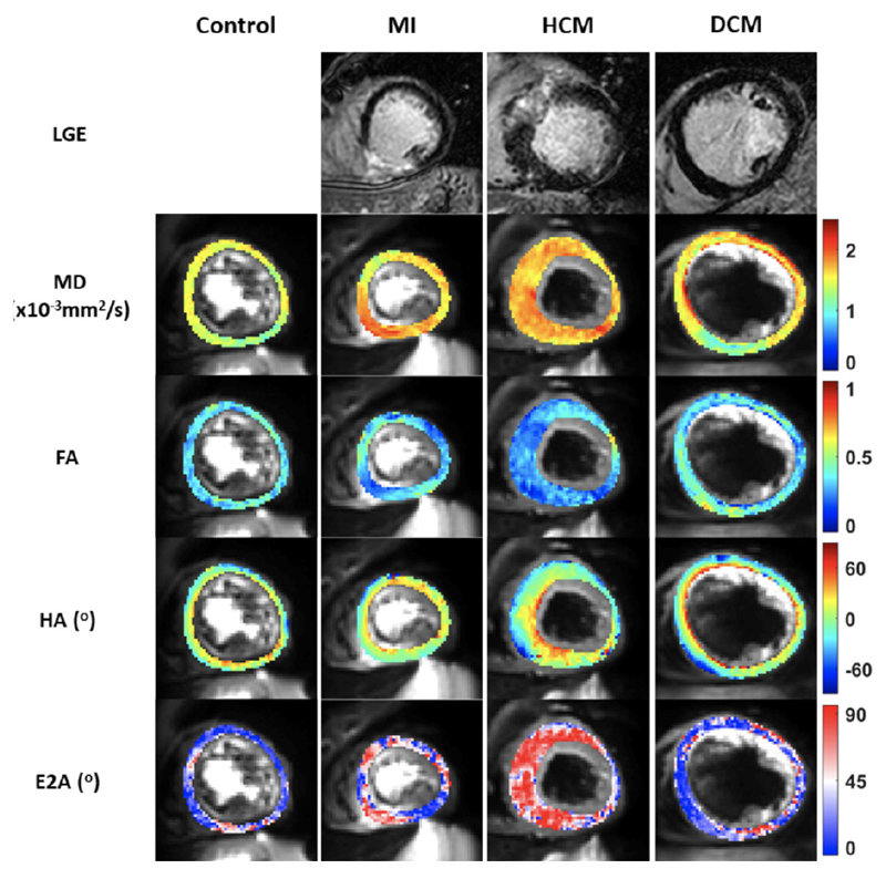
Figure 5.
Multiparametric imaging of the left ventricle myoarchitecture and disarray using diffusion tensor imaging (DTI) and late gadolinium enhancement (LGE). Column A displays a typical basal left ventricular (LV) slice obtained in a healthy control showing a normal mean diffusivity (MD), fractional anisotropy (FA), second eigenvector angle (E2A) and helix angle (HA). Column B demonstrates a basal LV slice in a patient with myocardial infarction (MI) with inferior LGE, raised MD and low FA. Column C demonstrates a basal LV slice in a patient with hypertrophic cardiomyopathy (HCM) with a raised MD and E2A and a low FA. Column D demonstrates a basal LV slice in a patient with dilated cardiomyopathy (DCM) demonstrating a raised MD and low FA and E2A.

