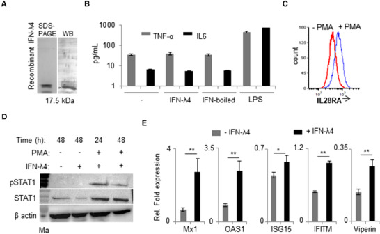
FIGURE 1.

IFN‐λ4 can signal in monocytes after PMA treatment. (A) Western blot analysis of the human recombinant IFN‐λ4 used in the study. (B) TNF‐α and IL‐6 cytokines secretion was measured by ELISA from cell‐free supernatants of THP‐1‐derived macrophage‐like cells treated for 48 h with 1 μg/ml IFN‐λ4, boiled IFN‐λ4 preparation, or 1 μg/ml of LPS; untreated macrophages served as control; mean and error bars depicting sd from technical replicates are shown. (C) THP‐1 cells were differentiated into macrophage‐like cells by treatment with PMA (100 nM) for 48 h. The cells were removed and compared with fresh untreated THP‐1 cells for surface expression of IL28RA using flow cytometry. (D) pSTAT1 and STAT1 expression was assessed by Western blot in THP‐1 cells treated or not with PMA (100 nM) in the presence or absence of IFN‐λ4 (1 μg/ml) for 24 and 48 h as indicated. β‐actin was used as loading control. The presented immunoblot is a representative image of two independent experiments. (E) IFN‐stimulated genes expression was analyzed by quantitative polymerase chain reaction (qPCR) based on RNA samples extracted from THP‐1‐derived macrophage‐like cells treated or not with 5 μg/ml of IFN‐λ4 for 24 h. The data are representative of two independent experiments showing mean of technical replicates from a single experiment with sd depicted by error bars. *P < 0.05; **P < 0.01
