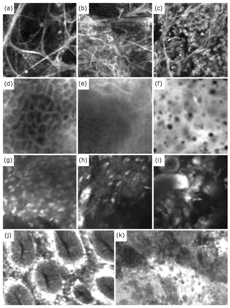
Fig. 4. Examples of structural changes observed in OEM images across variety of organ systems and conditions. These structural changes have been used to classify/detect a range of clinically relevant pathologies.
(a-c) Difference in tissue structure in the alveoli structures of the distal lung, indicating (a) healthy and (b) pathological elastin strands, as well as (c) alveoli sacs flooded with cells. (d-f) Difference between (d) healthy and (f) cancerous oral epithelium, along with (e) an example of oral epithelium with limited textural information where classification can be challenging. (g-i) Difference between (g-h) Glioblastoma and (i) Meningioma brain tumour images. (j-k) Difference between (j) healthy colon mucosa and (k) adenocarcinoma. Images (d-f) have been reproduced (cropped) from Figure 6 of the “Automatic Classification of Cancerous Tissue in Laserendomicroscopy Images of the Oral Cavity using Deep Learning” by Aubreville et al. (2017) under the Creative Commons Attribution (CC BY) 4.0 International License. Images (g-i) have been reproduced from Figure 3 of the “Automatic Tissue Differentiation Based on Confocal Endomicroscopic Images for Intraoperative Guidance in Neurosurgery” by Kamen et al. (2016) under CC BY 4.0. Images (j) and (k) have been reproduced (from Figures 2 and 3 respectively of the “Computer Aided Diagnosis for Confocal Laser Endomicroscopy in Advanced Colorectal Adenocarcinoma” by Ştefănescu et al. (2016) under CC BY 4.0 (https://creativecommons.org/licenses/by/4.0).

