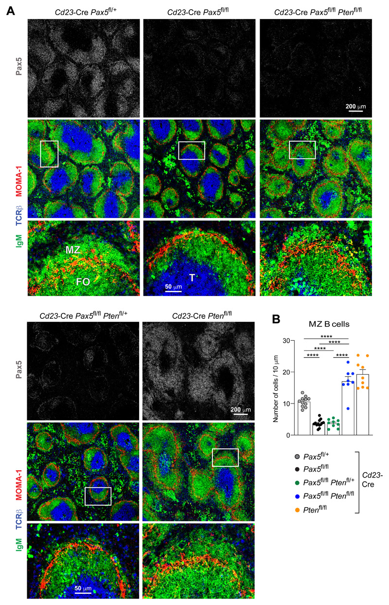
Figure 8. Rescue of MZ B cell development in Pten,Pax5 double-mutant mice.
(A) Immunohistological analysis of spleen sections from 12-week-old mice of the indicated genotypes. The sections were stained with antibodies detecting IgM (green), MOMA-1 (red), TCRβ (blue) and Pax5 (gray). Selected areas (boxed) of B cell follicles are shown at higher magnification. T, FO B and MZ B cell zones are indicated. One of three experiments is shown. (B) Quantification of the MZ B cells on histological sections. The average number of IgM+ B cells outside of the MOMA-1+ macrophage ring was determined per 10 μm length of the perimeter of the MOMA-1+ ring (see Methods). Each dot represents the measurement of one follicle. The mean values determined for the indicated genotypes is shown with SEM and were analyzed by one-way ANOVA with Tukey’s multiple comparison test; ****P < 0.0001.

