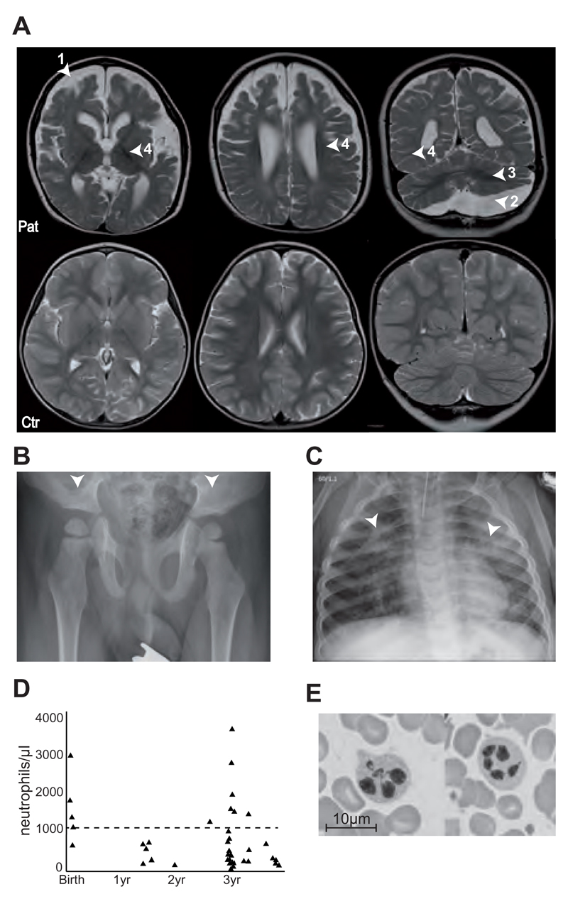
Figure 1. Clinical phenotype of the patient.
(A) MRI scan of the brain at 3.5 years (upper panel), showing atrophy of the telencephalon (1), enlarged external and internal cerebrospinal fluid spaces (4), arachnoidal cyst (2) in the posterior fossa and insufficient myelination (3) indicated by arrow heads. The lower panel shows an MRI scan of a healthy 3.5 year old child. (B) X-ray of the pelvis showing flat, dyplastic acetabulae. (C) X-rays of the chest showing chronic interstitial pneumonia. (D) Neutrophil counts over time. Dashed line at 1000 neutrophiles/μl represent limit for neutropenia. (E) Bone marrow smear showing hypersegmented neutrophils.

