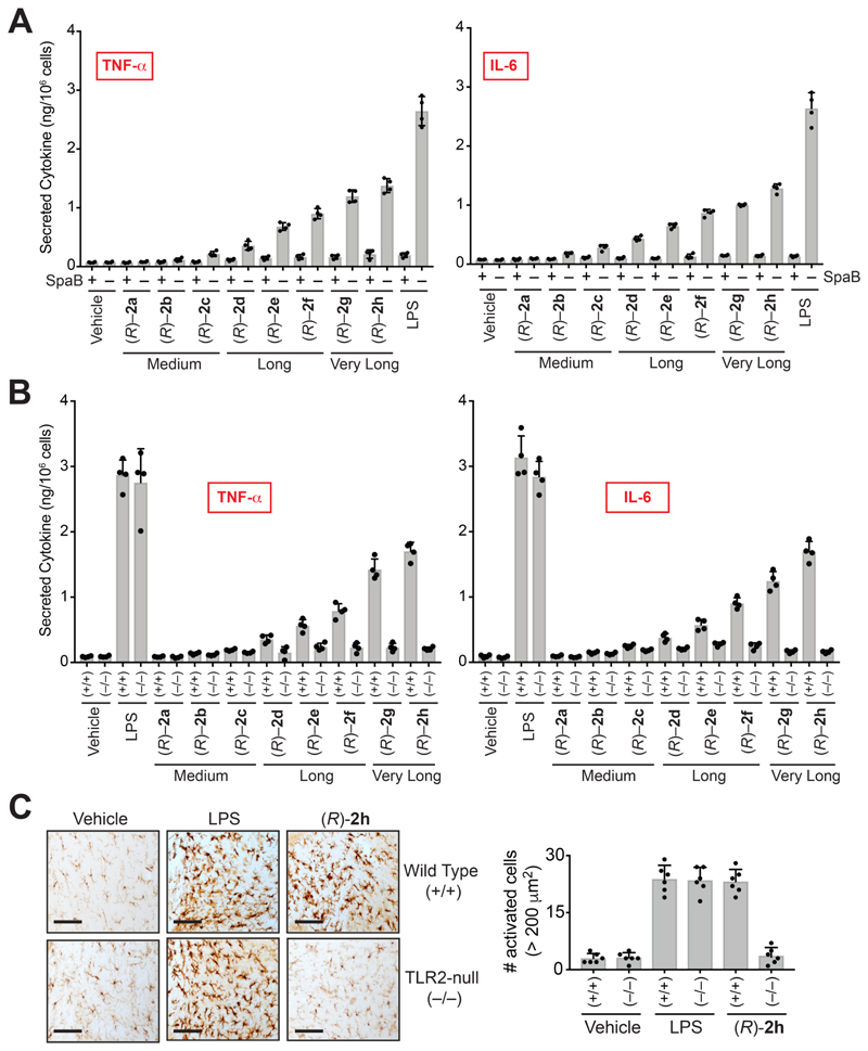
Figure 3. VLC lyso-PSs elicit pro-inflammatory responses via a TLR2 dependent pathway.
Secreted TNF-α and IL-6 from PPM harvested from (A) wild type mice pre-treated with DMSO or SpaB (10 㭜M, 4 hours, 37 °C), followed by treatment with vehicle or LPS or (R)-2a-h (1 㭜M, 4 hours, 37 °C); (B) wild type (+/+) or TLR2 knockout (–/–) mice following treatment with vehicle or LPS or VLC (R)-Me-lyso-PSs (1 㭜M, 4 hours, 37 °C). (C) Representative images from Iba-1 immunostaining for microglial activation (black bar = 250 㭜m) and quantification of enlarged cells (>200 㭜m2) in the cerebellum (per 1.44 mm2) of wild type (+/+) or TLR2 knockout (–/–) mice following intravenous injection of vehicle (PBS), LPS or (R)-2h (C24:0) (all 1 mg/kg body weight, 10 hours). All data presented in (A), (B), and (C) is represented as mean ± standard deviation (n = 4-6/group).

