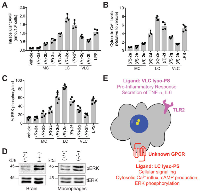
Figure 4. LC lyso-PSs activate macrophages through a putative GPCR.
(A) Intracellular cAMP, (B) relative cytosolic Ca2+ levels, and (C) percentage of phosphorylated ERK from WT PPM following treatment with vehicle (DMSO) or LPS or (R)-2a-h (1 㭜M, 10 mins, 37 °C). All data presented in (A), (B), and (C) is represented as mean ± standard deviation (n = 4-5/group), where MC = medium chain, LC = long chain and VLC = very-long chain. (D) Representative western blots on lysates of brain (6-month-old mice) and LPS-treated (1 㭜M, 4 hours, 37 °C) PPM harvested from wild type (+/+) or ABHD12-null (–/–) mice, showing enhanced phosphorylation of ERK in both these tissues for the ABHD12-null (–/–) genotype. (E) Schematic cartoon representation summarizing the lyso-PS signaling pathways by possibly two types of receptors on mammalian macrophages.

