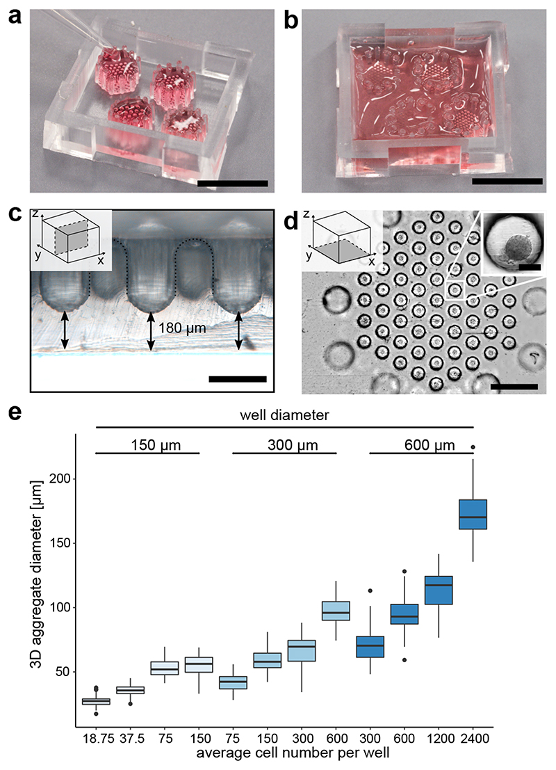
Fig. 1. Microwell chips for generating and culturing 3D cell aggregates.
a, Image of the microwell chip with four microwell arrays (top-down view). Each hexagonal array is surrounded by 12 pillars to keep fluid volume above the microwell. Cells were seeded in 20-40 μL. Scale bar is 1 cm. b, After cell seeding, the entire microwell chip was filled up to 800 μL media for long-term cell culture. Scale bar is 1 cm. c, Cross-sectional view of the microwell chip bottom. Scale bar is 300 μm. d, Bottom view on one microwell array. Scale bar is 1 mm. Inset: Higher magnification of a pancreatic-progenitor-derived 3D aggregate formed from 600 cells. Scale bar is 100 μm. e, Aggregate size in dependence on the cell number and well diameter after 24 hours of seeding pancreatic progenitors, including at least 58 pancreatic-progenitor-derived 3D aggregates from three different microwell arrays. Boxplots display the median with the first and third quartile, whiskers denote the 1.5x interquartile range and outliers are marked as dots.

