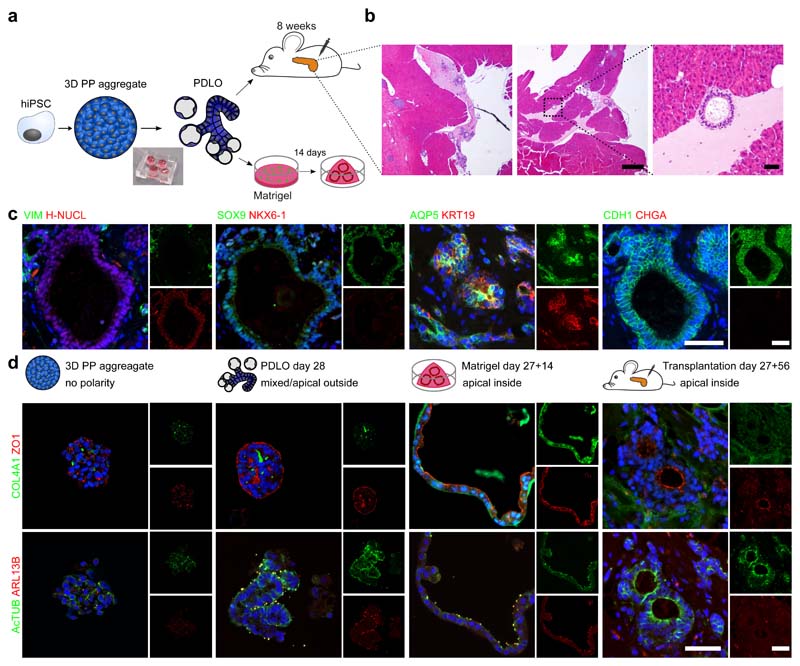
Fig. 3. Apical-out polarity of the microwell-chip-derived PDLOs switched upon orthotopic transplantation or embedding into Matrigel.
a, Schematic of the Matrigel and orthotopic transplantation experiment. PDLOs were transplanted on day 27 and mice were sacrificed after 8 weeks. b, Overview haematoxylin-eosin (HE) staining and magnification of the engraftment site depicted by the dashed square (n=2 mice). Scale bar: 500 μm for overviews, 50 μm for magnification. c, PDLOs formed human epithelial duct-like tissue in vivo. (H-NUCL: human-specific nucleoli). Scale bar denotes 50 μm. d, IF images for the apical markers ZO1 and AcTUB and basal markers COL4A1 and ARL13B on 3D pancreatic-progenitor aggregates, PDLOs, Matrigel PDLOs and transplanted PDLOs. Complementary images are depicted in Supplementary Fig. 5. The nuclei were counterstained with DAPI. Scale bar denotes 50 μm.

