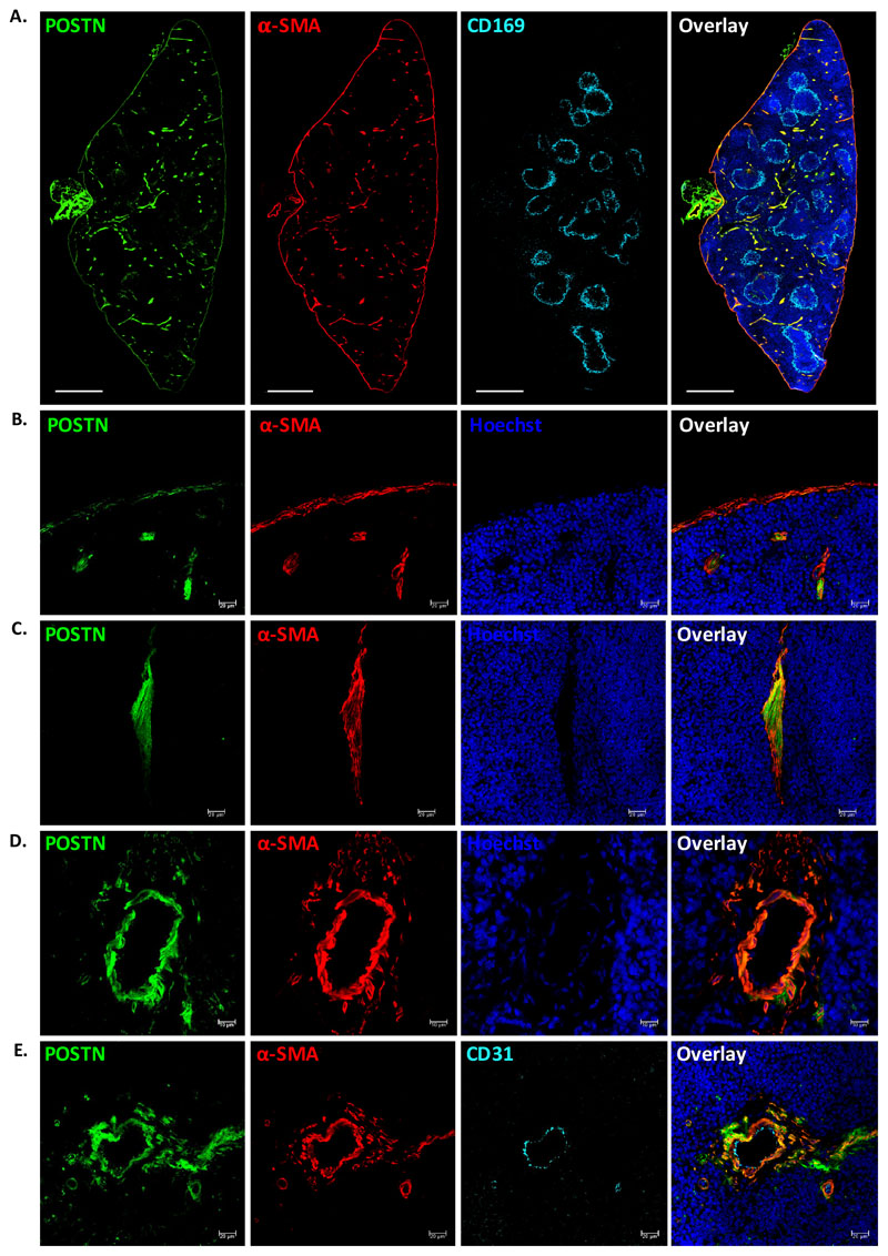
Figure 1. POSTN is expressed by myofibroblasts in adult spleen.
(A) POSTN expression in adult spleen tissues was examined using immunohistochemistry on 10μm cryo-sections. WP areas were identified by using marginal zone macrophage marker CD169; trabecular, capsular and vascular regions were identified by using antibodies against α-SMA. Specific antibodies were used to identify the cells expressing POSTN, nuclear counterstaining was done using Hoechst 33342. The sections were visualized and tile scan was performed on confocal microscopes. (n=2, N=8, scale bar=0.5mm).
(B-D) Regions with compelling levels of POSTN expression; namely, the capsule (B), trabeculae (C), and vasculature (D) were analyzed by immunostaining with myofibroblast/smooth muscle cell marker α-SMA. (n=5, scale bar= 20μm for panel B,C and 10μm for panel D).
(E) The endothelial and smooth muscle lining of the vessels were identified using antibodies against endothelial marker CD31 and α-SMA, along with anti-POSTN antibody (n=3, scale bar=20μm).

