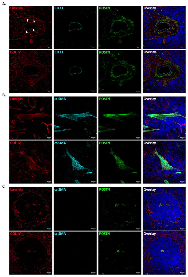
Figure 2. POSTN colocalizes with ECM proteins close to myofibroblasts.
Immunostaining experiments were performed to examine colocalization of POSTN with ECM proteins laminin and collagen IV in various anatomical locations within spleen tissue. (A) Vascular region of spleen identified by CD31 immunostaining. Colocalization of POSTN with laminin (upper panel) and collagen IV (lower panel) visualized using specific antibodies. (B) Staining for α-SMA was used to identify trabecular area, wherein POSTN colocalization with ECM was examined by counterstaining with laminin (upper panel) and collagen IV (lower panel). (C) White pulp area, identified by the laminin (upper panel) and collagen IV (lower panel) expression on the periphery and in red pulp. Identification of smooth muscle cells/myofibroblasts and POSTN using specific antibodies.
n=3, Scale bar=20μm

