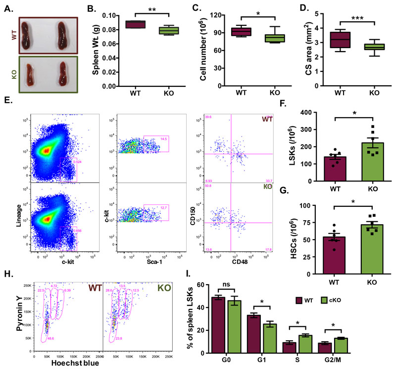
Figure 5. Loss of POSTN expression affects proliferation of hematopoietic progenitors in spleen.
(A) Spleen tissues from Postn+/+ (WT) and Postn-/- (KO) mice were harvested. (B) Comparison of whole spleen weight from WT and KO mice. (C) The total number of MNCs from spleens of WT and KO mice. (D) Cross-sectional area quantified using transverse sections of spleen tissues from WT and KO mice. The formalin-fixed, paraffin-embedded tissues were used to cut 10μm sections that were used for H&E staining and analysis. (E-G) Quantification of frequency of various hematopoietic stem and progenitor cell populations in the spleen from WT and KO mice; (F) LSK cells, (G) primitive HSCs. (H) Cell cycle analysis of the spleen LSK cells. Total MNCs from spleen tissues were used to first label the cells with lineage, Sca-1 and c-kit antibodies followed by Hoechst 33342/Pyronin Y staining. Samples acquired on a flow cytometer were analyzed and LSK cells were gated for further analysis for Hoechst 33342 and Pyronin Y intensity. (I) Comparison of the proportion of spleen LSK cells in various stages of cell cycle (n=6).
Unpaired two-tailed Student’s t-test was performed. * p<0.05, ** p<0.01, ***p<0.001. ns indicates not significant.

