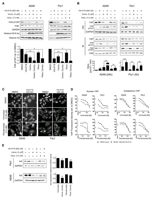
Figure 4. Combined inhibition of HDAC and FAK abolishes FAK kinase activity and regulates YAP localization.
A, Lysates from A549 (left) and Flo1 (right) cells treated as indicated were immunoblotted for FAK, phosphorylated FAK (Y397), GAPDH, histone H3, and acetylated (K-Ac) histone H3. Quantification of phosphorylated FAK (Y397) immunoblotting (bottom) is displayed as means ± SEM (n = 3 independent experiments). *, P < 0.05; **, P < 0.01 (one-way ANOVA). B, Lysates from A549 (left) and Flo1 (right) cells were immunoblotted for YAP, phosphorylated YAP (S127), FAK, phosphorylated FAK (Y397), and GAPDH at 5 and 24 hours post drug treatment. C, A549 (left) and Flo1 (right) cells were fixed and labeled with anti-YAP antibody after 24 hours of drug treatment. Scale bar, 50 μm. D, Quantification of nuclear and cytoplasmic anti-YAP labeling. Relative fluorescence intensity values of anti-YAP labeling in the nuclear (left) and cytoplasmic (right) cellular compartments were normalized to DMSO values and displayed as means ± SEM (n = 3 independent experiments). E, Lysates from Flo1 (top) and A549 (bottom) cells treated as indicated were immunoblotted for Axl and GAPDH. Quantification of Axl expression is displayed as means ± SEM (n = 3 independent experiments). *, P < 0.05; **, P < 0.01 (one-way ANOVA).

