Figure 2.
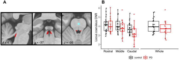
Locus coeruleus imaging. (A) Study specific locus coeruleus atlas, also showing the reference region (light blue) in the central pons. (B) CNR for the locus coeruleus subdivisions and whole structure in Parkinson’s disease patients (PD) versus controls (note, left and right locus coeruleus are combined).

