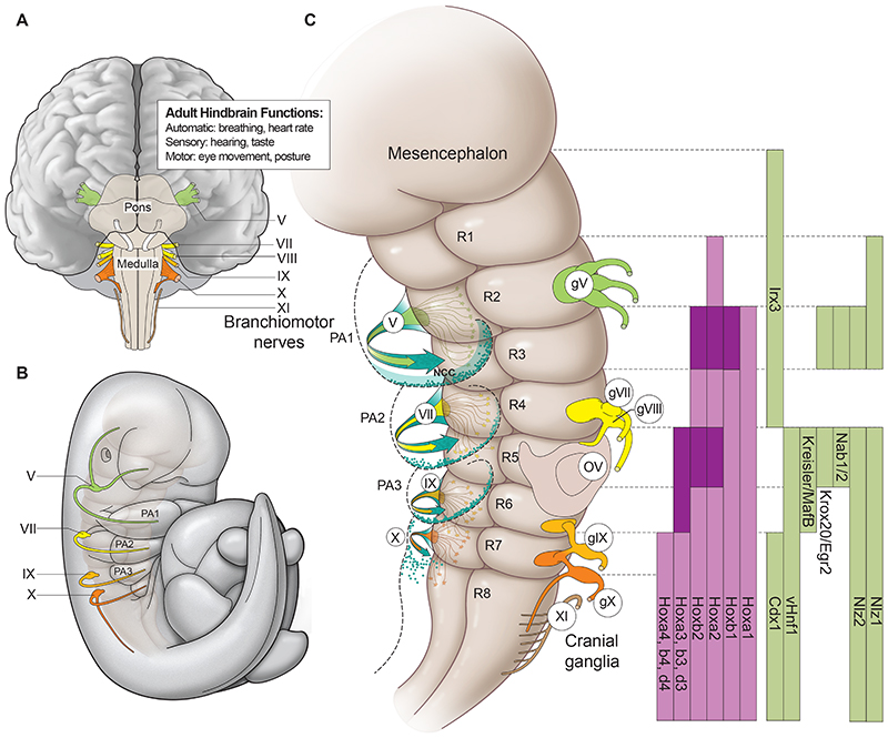
Fig. 1. Anatomy and segmental organisation of the hindbrain.
(A) Diagram of ventral aspect of an adult mouse hindbrain noting the relative positions of branchiomotor nerves and several of its functional roles. (B) Drawing of a lateral view of an E10 mouse embryo showing the paths of branchiomotor nerves and their relationship to the pharyngeal arches (PA). (C) Diagram of a ventral view of a developing hindbrain depicting individual rhombomeres (R) and their relationship with the positions of cranial ganglia (gV-X1), motor nerves (V-X), migrating streams of cranial neural crest cells (NCC) and pharyngeal arches. There is a two-segment periodicity in the segmental organisation, as cranial ganglia, and major streams of migrating neural crest cells (blue arrows and dots) are associated with even-numbered rhombomeres. Each branchiomotor nerve is derived from alternating pairs of even- and odd-numbered segments, exiting to the periphery from the dorsal aspects of only the even-numbered segments. Rhombomere-restricted domains of gene expression of key transcription factors involved in segmentation and segmental patterning are indicated on the right. The gene names correspond to mouse and may vary in other vertebrates. Darker shades of colour (purple, Hox genes; green segmental subdivision genes) in the expression domains indicate higher levels of expression in specific rhombomeres.

