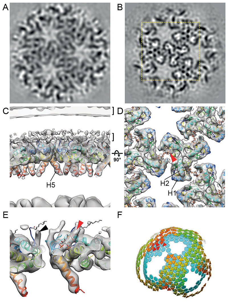
Fig. 3. The mature HIV-1 matrix structure.
(A, B) Slices through reconstructions of the mature HIV-1 MA lattice in cHIV (A) and cHIV MA-SP1 (B). The boxed region in (B) is shown at a higher magnification in (D). (C, D) An isosurface view of the cryo-ET reconstruction for mature cHIV MA-SP1 MA lattice fitted with the structure of monomeric MA, cut perpendicular to the membrane (C) or viewed from the top towards the virus center (D). The two layers of density corresponding to the lipid headgroup layers are indicated by brackets. The structure of monomeric MA determined by NMR (PDBID: 2H3Q (18); colored blue to red from N to C terminus) was fitted as a rigid body into the density. Helices 1, 2 and 5 are marked. Density is observed in the PI(4,5)P2 binding site (red arrowhead), and the structure of PI(4,5)P2 as resolved bound to MA by NMR (PDBID: 2H3V (18)), is shown as a stick model. (E) As in (C), enlarged and cut to reveal density corresponding to the N-terminal residues (black arrowhead), and PI(4,5)P2 (red arrowhead). (F) As in Fig. 2G, lattice map for the mature cHIV MA-SP1 MA lattice and the underlying CA lattice.

