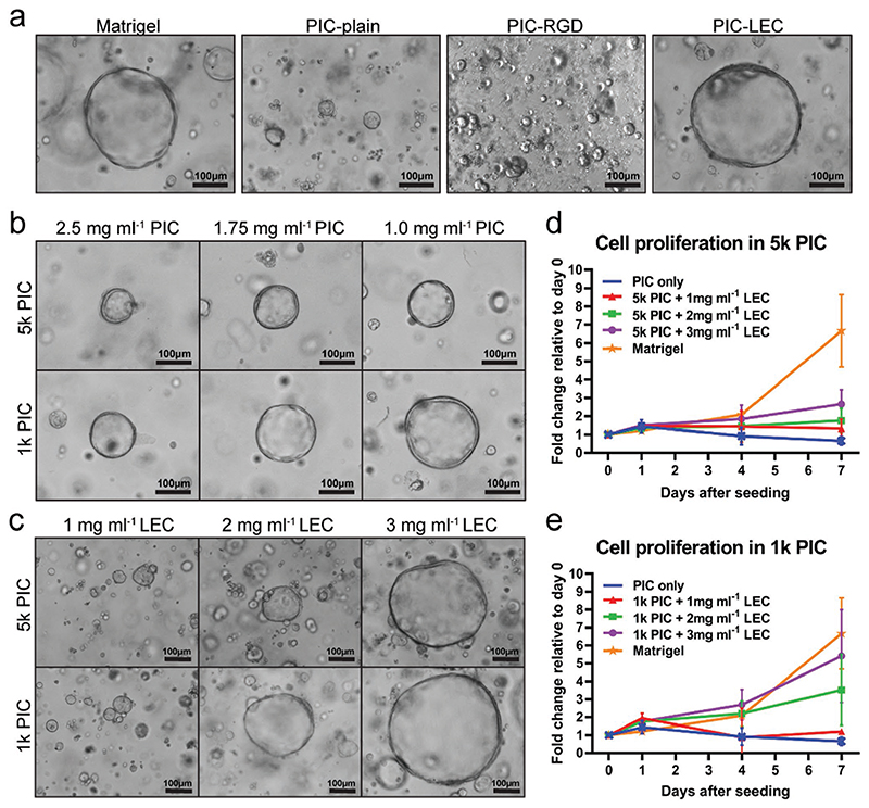
Figure 1. An optimized PIC hydrogel supports liver organoid expansion.
Single organoid cells were seeded at day 0 and cultured in human organoid expansion medium (EM) supplemented with the Rho kinase inhibitor Y-27632 for 14 days. Four different donors were analyzed in independent experiments (N = 4). a) Light microscopy images of organoids at day 7 after single cell seeding. Organoids did not proliferate in PIC-plain and PIC-RGD, but showed a morphology comparable to Matrigel in PIC supplemented with 3 mg mL−1 LEC (PIC-LEC). b) Lower concentrations of PIC improve organoid proliferation. c) Light microscopy pictures reveal a dose-dependent effect of LEC on organoid proliferation. Quantification of organoid proliferation in d) 5k PIC and e) 1k PIC. Organoids from five different donors were cultured in Matrigel or PIC with different concentrations of LEC. Relative cell numbers were determined by an Alamar blue assay every 2-3 days and cell expansion relative to day 0 was calculated. Each dot represents the mean of the four donors with standard deviation.

