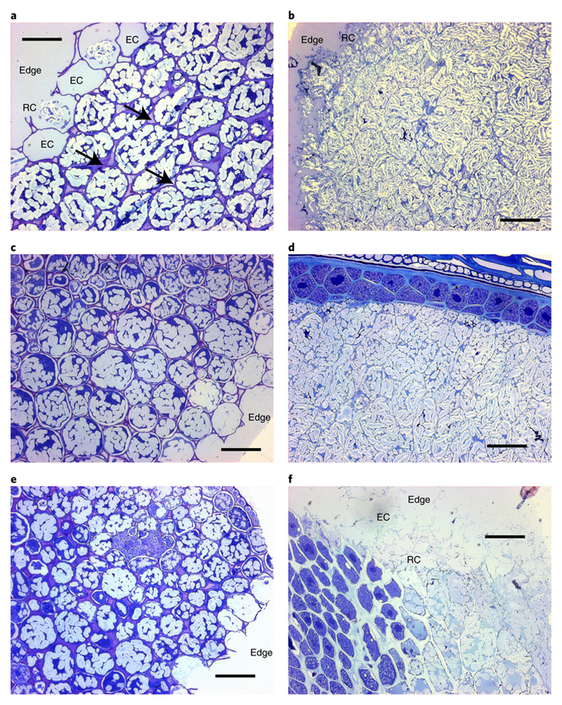
Figure 2. Microstructure of hydrothermally-cooked intact tissue macroparticles.
Cross-sections of chickpea (left, a,c,e) and wheat (right, b,d,f), before (a,b), and after (c,d,e,f) digestion. Light micrographs of cross-sections of chickpea (left, a,c,e) and wheat (right, b,d,f) cut to 0.5 μm thickness and stained with toluidine blue (1% w/v, with 1% w/v sodium borate). Scalebar = 50 μm. In micrographs captured before digestion (a,b), the cell walls are seen to surround intracellular starch within the intact tissue, with some ruptured (‘RC’) and/or empty (‘EC’) cells present on the particle edges (i.e., the fractured surface created by dry-milling). Arrows indicate some of the areas where weakening of inter-cellular linkages has occurred. The internal structure and edges of chickpea tissue examined after 4 h of in vitro digestion (c) did not appear to be altered. After 2 h digestion, wheat starch was still evident within many endosperm cells, particularly those in close proximity to the aleurone layer or crease (d). The appearance of chickpea tissue remained unchanged after 6 h (e), whereas wheat endosperm cells near the particle edges had collapsed and/or had been eroded (‘edge’) after 6 h (f).

