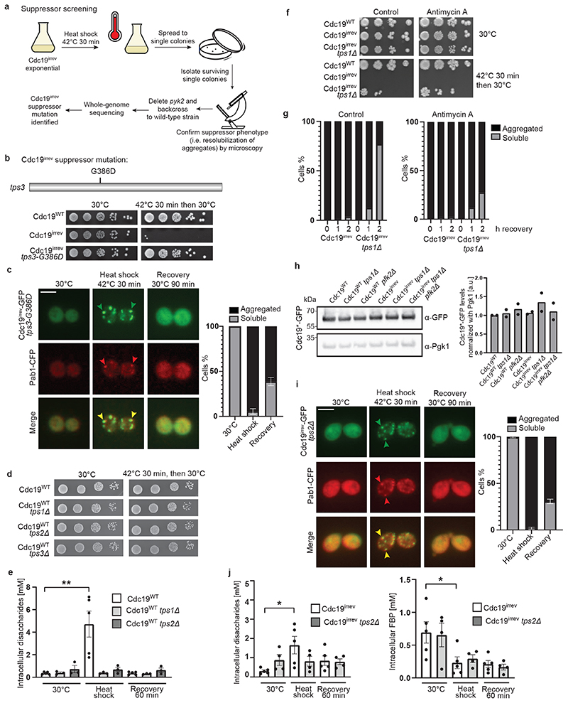
Extended Data Fig. 2. Genetic screening identifies trehalose metabolism as a regulator of reversible Cdc19 aggregation.
(A) - (D) Screening protocol that identified a single point mutation (G386D) in TPS3 as a suppressor of stress-induced growth arrest of cdc19irrev cells (A). Serial dilutions of the indicated strains were spotted on agar plates before or after heat shock (42 °C, 30 min), and imaged after 3 days at 30 °C (3 independent experiments) (B and D). tps3-G385D cells expressing Cdc19irrev-GFP and Pab1-CFP were heat shocked (42 °C, 30 min) and allowed to recover at 30 °C (C). Plot indicates mean percentage (%) of cells with Cdc19 aggregates ± S.E.M (n = 3 independent experiments, >30 cells per sample/experiment). Scale bar: 5 μm.
(E) Intracellular disaccharides were measured in the indicated strains before, during and after heat shock (42 °C, 30 min), and shown as mean ± S.E.M. (n = 5 independent experiments for wild-type, n = 3 independent experiments for tps1Δ and tps2Δ, two-tailed Mann-Whitney test, P = 0.0079).
(F) and (G) The indicated strains were heat shocked (42 °C, 30 min) and allowed to recover at 30 °C ± antimycin A (1 or 2 μM, respectively). (F) Serial dilutions were spotted on agar plates ± antimycin A and imaged after 3 days at 30 °C (3 independent experiments). (G) Plots indicate mean percentage (%) of cells that re-solubilized Cdc19 from two independent experiments.
(H) Mean Cdc19-GFP levels relative to Pgk1 in the indicated strains are shown from two independent experiments.
(I) tps2Δ cells expressing Cdc19irrev-GFP and Pab1-CFP were heat shocked (42 °C, 30 min) and allowed to recover at 30 °C. Plot indicates mean percentage (%) of cells with Cdc19 aggregates ± S.E.M (n = 3 independent experiments, >30 cells per sample/experiment). Scale bar: 5 μm.
(J) Intracellular disaccharides (mainly trehalose [46]) and FBP were measured in the indicated strains before, during and after heat shock (42 °C, 30 min), and plotted as mean ± S.E.M. (n = 4 independent experiments for cdc19irrev tps2Δ, n = 5 for cdc19irrev, two-tailed Mann-Whitney test, PDisaccharides = 0.0159, PFBP = 0.0317). Source data for all graphical representations and unprocessed Western blots available in Source Data Extended Data Fig. 2.

