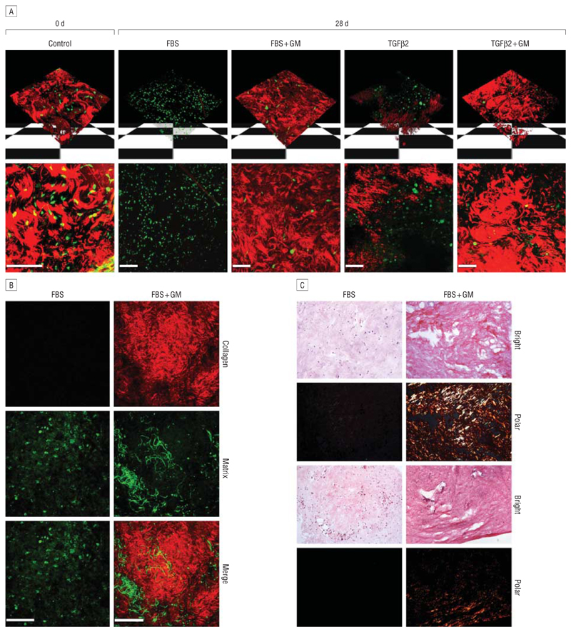
Figure 7.
Matrix metalloproteinase inhibitor GM6001 (GM) prevents collagen fiber breakdown in porcine conjunctiva. A, The upper panel shows 3-dimensional reconstruction of z-stack confocal images, and the lower panel shows a flat projection of all sections (red, matrix; green, live cells). In fresh tissue (Control), thick masses of collagen fibers can clearly be seen. After 4 weeks in FBS or TGFβ2, fiber degradation is observed, which is significantly reduced in the presence of GM6001. Viable cells are present in the segments in all conditions. B, Second harmonic generation shows that FBS degrades collagen fibers (red), which is prevented in the presence of GM. The green panel represents the 2-photon autofluorescence of the sample, showing cells and other fibers of the connective tissue. C, Picrosirius red staining of porcine tissue samples after 4 weeks in culture. Brightfield images were obtained (Bright), showing total collagen (red-pink) in the tissue. A polarizing filter (Polar) was also used to monitor the birefringence of the thick collagen fibers (orange-yellow), which are detectable in samples that have been maintained in GM but are undetectable in those without GM. Shown are 2 representative fields for each condition. Scale bar, 100 μm.

