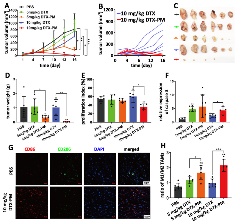
Figure 3. Inhibition of SGC7901 cell line-derived subcutaneous xenografts by DTX-PM.
A: Tumor growth curves of mice treated 5 times with PBS, DTX and DTX-PM at 5 and 10 mg/kg, showing that DTX-PM were more effective than free DTX. B-D: DTX-PM dosed at 10 mg/kg was able to completely eradicate tumors in almost all mice. E and F: Proliferation and apoptosis assessed by immunohistochemical staining Ki67 and cleaved caspase 3. G: The polarization of tumor-associated macrophages (TAM) was studied by immunofluorescence staining. H: Calculation of the ratio between M1-like macrophages (CD86 staining; red) and M2-like TAM (CD206 staining; green) showed that significantly more M1-like TAM were present upon treatment with DTX-PM. Values are mean ± SD. *: P < 0.05, **: P < 0.01, and ***: P < 0.001.

