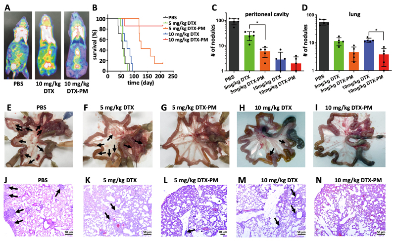
Figure 4. PM improve DTX efficacy in metastatic CDX models of GI cancer.
A: 18F-FDG-based PET-CT imaging of peritoneal metastasis upon treatment with PBS, 10 mg/kg DTX and 10 mg/kg DTX-PM at day 16. B: Survival of mice with peritoneal GI cancer metastasis upon five administrations of PBS, DTX and DTX-PM at 5 and 10 mg/kg. C-N: Assessment of metastatic nodules in the peritoneal cavity (C) and in the lungs (D), based on macroscopic quantifications in the intestines of mice (E-I) and on microscopic analyses of the lungs of mice (J-N). Metastatic nodules are indicated by black arrows. Values represent mean ± SD. *: P < 0.05, **: P < 0.01, and ***: P < 0.001.

