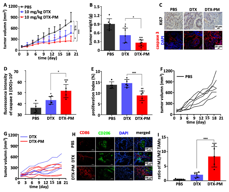
Figure 5. DTX-PM improve DTX efficacy in a PDX model of GI cancer.
A: Tumor growth inhibition upon treatment with PBS, and with 10 mg/kg of free DTX and DTX-PM. B: Tumor weights at the end of the experiment, showing significantly more efficient tumor inhibition for DTX-PM than for free DTX. C-E: Immunofluorescence analysis of cellular proliferation (Ki67) and apoptosis induction (cleaved caspase 3), confirming that DTX-PM was more effective than free DTX. F and G: Individual PDX tumor growth curves in the mice treated with DTX and DTX-PM as compared to PBS controls, exemplifying the superior efficacy of DTX-PM. H and I: PDX tumors were analyzed by means of immunofluorescence microscopy, showing a substantially increased ratio of M1-like to M2-like TAM upon DTX-PM treatment.

