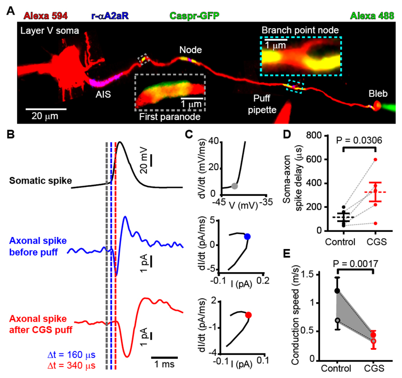
Fig. 4. A2a receptors in the node of Ranvier modulate conduction velocity.
(A) Myelinated axon in Thy1-Caspr-GFP mouse filled with Alexa 594 and patch-clamped at the cell soma and end-of-axon bleb. (B) Average of >100 evoked action potentials in the soma and bleb in control conditions and while puffing 0.5 μM CGS 21680 at a node of Ranvier. Dashed lines show times of initiation of action potential derived from threshold values of dV/dt and dI/dt (see text). (C) Phase plane plots showing times indicated on B (dots). (D) Response latency in bleb. (E) Conduction velocities derived making assumptions discussed in the main text (closed circles assume spike starts at the middle of the AIS and the forward speed is twice the backward speed; open circles assume spike starts at the end of the AIS and the forward speed is three times faster than the backward speed). Data in A-C are from mouse; data in D-E combine data from rats and mice (neither the initial speed nor the percentage change evoked by CGS 21680 differed significantly between rats and mice, p=0.43 and 0.15 respectively).

