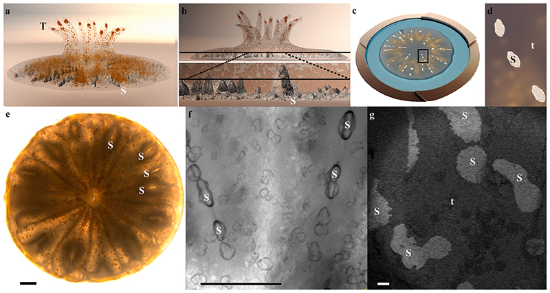
Fig. 1. A primary polyp and its forming mineralized septa.
(a) Illustration of a few days old primary polyp. Endosymbionts are orange, and mineral is grey. (b) Upper panel: An illustration of the same polyp as in (a) in a side view, with internal plane revealed by cryo-planing or freeze–fracture marked with a black line. Bottom panel: Magnification of this plane crossing two septa. (c) Illustration of high-pressure frozen, cryo–planed primary polyp (top view) vitrified in natural seawater (blue) inside a high-pressure freezing disc (gold). (d) Magnification of the area marked with black rectangle in (c). (e) Light microscopy image of a primary polyp. (f) Higher magnification wide-field microscope image of two forming septa. (g) Cryo-SEM (EsB mode) micrograph of freeze fractured primary polyp showing the non-continuous fracture surface of two forming septa. Septa mineral surface appears white, and coral tissue surface appears grey. T-tentacles, S-septum mineral, t-tissue. Scale bars: e, f, – 100 μm, g – 20 μm.

