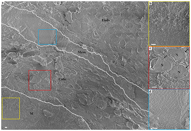
Fig. 2. A cryo-SEM micrograph of aboral body layers observed in a high–pressure frozen, freeze-fractured primary polyp.
(a) An overview image showing aboral body layers depicted by white separating lines including the mineral, M, (Yellow rectangle is magnified in (b)), the calicoblastic cell layer, Calic, (Red rectangle is magnified in (c)), the noncellular mesoglea, Meso, (Light blue rectangle is magnified in (d)) and the endoderm (Endo). N-Nucleus. All scale bars are 1 μm.

