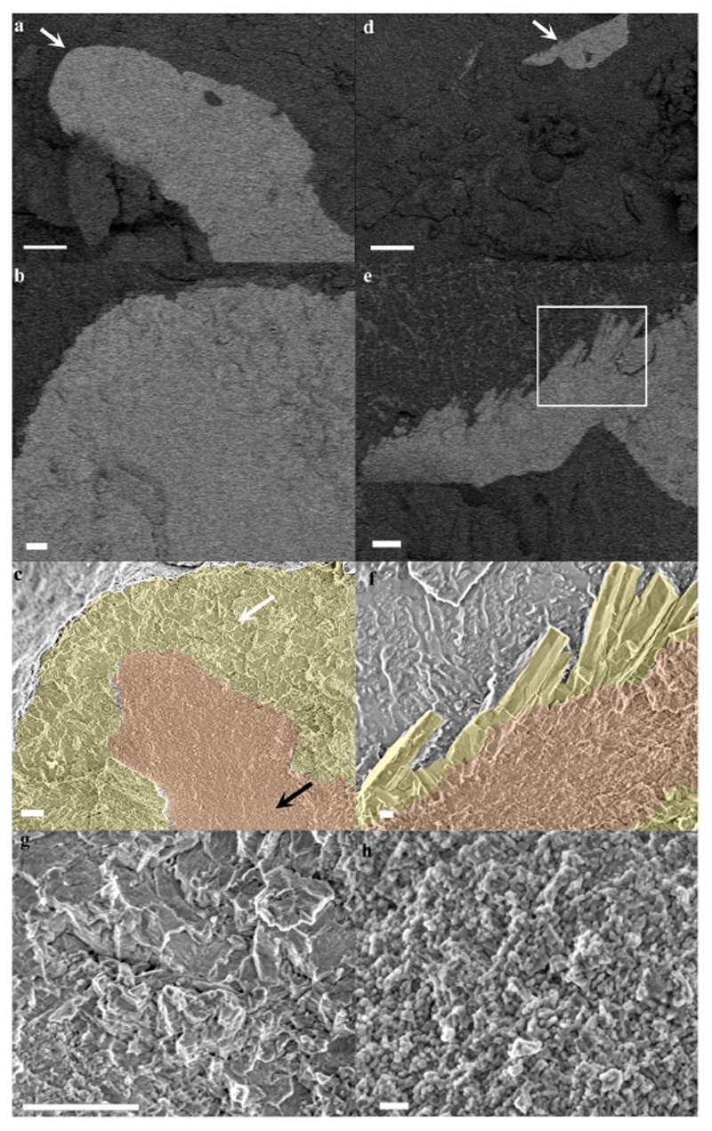
Fig. 3. Cryo-SEM micrographs of mature and forming septa in a primary polyp.
(a) A mature septum imaged in EsB mode. Mineral appears brighter than its surrounding tissue. (b) Magnification of the area pointed with white arrow in (a). (c) The same areas as in (b) imaged in SE mode with CoC and elongated microcrystals highlighted by false coloring. (d) A newly formed septum imaged in EsB mode. (e) Magnification of the area pointed with white arrow in (d). (f) SE mode image of the area marked with white rectangle in (e) with CoC and elongated microcrystals highlighted by false coloring. (g) Higher magnification of micron sized crystals’ fracture surface pointed with white arrow in (c). (h) Higher magnification of CoC nano-spheres texture pointed with black arrow in (c). Orange-CoC. Yellow-elongated microcrystals. Scale bars are (a)-and (d)-10 μm, (b), (c), (e) and (g)-1 μm, (f)-200 nm, (h)-100 nm.

