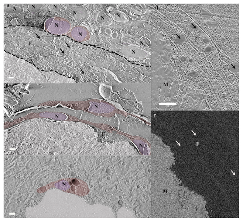
Fig. 4. Calicoblastic cell morphologies observed in freeze-fractured (a-b) and cryo-planed (c-e) primary polyps using cryo-SEM.
(a) Calicoblastic cell layer (imaged locus is the same as in Fig. 2c with the septum mineral and two representative calicoblastic cells highlighted by false coloring. A filopodia network found between the calicoblastic cell bodies and the septum is denoted between two dashed black lines. Four representative vesicles contained within the filopodia network are denoted with black arrows. (b) Three elongated calicoblastic cells found in close proximity with the mineral. (c) A cup shaped calicoblastic cell attached to the mineral surface. (d) High magnification of filopodia found in close proximity with the mineral (SE mode). (e) The same field of view as in (d) imaged in EsB mode. False coloring is used to highlight: cell nucleolus (pseudo-purple), cell body (pseudo-red), septum mineral (pseudo-white). Scale bars are 1 μm. N-nucleus, F-filopodia, M-mineral.

