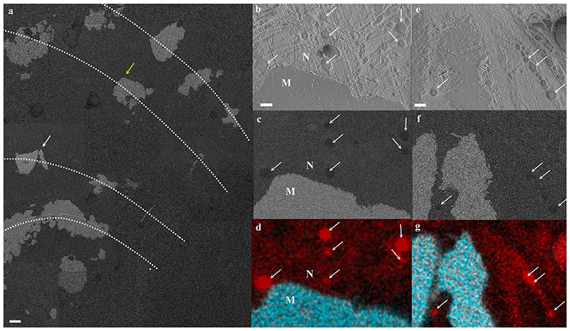
Fig. 5. Cryo-SEM/EDS analysis of calicoblastic cells in a cryo-planed primary polyp.
(a) An overview collage image composed of several high magnification cryo-SEM (EsB mode) micrographs stitched together, showing the cryo-planed surface along several septa (dotted lines represent septa long axes) in a primary polyp. The mineral surfaces of the planed septa appear white, and the coral soft tissues appear grey. (b-d) High magnification of locus pointed with a yellow arrow in (a) (same area as in Fig. 4c showing a cup-shaped calicoblast attached to the mineral imaged in SE, EsB and EDS map, respectively. (e-g) High magnification of locus pointed with a white arrow in (a) showing filopodia in close proximity with the mineral imaged in SE, EsB, and EDS maps, respectively. Carbon-rich vesicles are pointed with white arrows in (b)-(g). EDS maps show carbon (red) and calcium (turquoise) distributions. M-mineral, N-nucleus. Scale bars: (a)-10 μm, (b-g)-1 μm.

